Diagram Of Animal Cell Under Electron Microscope / Difference Between Plant And Animal Cells Cells As The Basic Units Of Life Siyavula / Resolving power is the ability to distinguish between separate things which are close to each other.
byMitchel Schamberger-0
Diagram Of Animal Cell Under Electron Microscope / Difference Between Plant And Animal Cells Cells As The Basic Units Of Life Siyavula / Resolving power is the ability to distinguish between separate things which are close to each other.. Transmission electron microscopy is a proven technique in the field of cell biology and a very the most common routes are indicated in the following schematic diagrams: Ishita observed a slide of eukaryotic cell under electron microscope. We say cells are microscopic because they can only be seen under a microscope. Here's a diagram of a plant cell: Ppt structure of plant and animal cells under an electron.
The diagram shows part of a cell surface membrane. Cell scanning electron microscope hd stock video 717 725 243. Cell structure teaching resources the science teacher, organelles biology for majors i, 11 different types of cells in the human body, class test, chronic inflammation under the microscope learn share. Animal cell organelles and functions with diagrams. The diagram shows a stage micrometer, with 23.
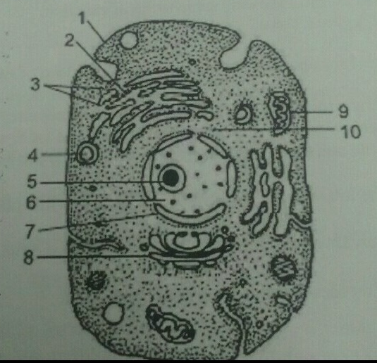
Plzz Answer This Q Q 19 Given Below Is A Diagrammatic Sketch Of Electron Microscopic View Of An Animal Science The Fundamental Unit Of Life 12392108 Meritnation Com from s3mn.mnimgs.com The diagram shows a phospholipid bilayer (cell membrane) with carbon dioxide molecules on one side of. Resolving power is the ability to distinguish between separate things which are close to each other. Typical animal cell pinocytotic vesicle lysosome golgi vesicles golgi vesicles rough er (endoplasmic reticulum) smooth er (no ribosomes) cell (plasma) 2. Preparing samples and using the electron when you look at animal or plant cells under the electron microscope, you can see a lot more detail. Plant cells have cell walls, one large vacuole per cell, and chloroplasts, while animal cells will have a cell membrane only. All living things are made up of one or more cells the cell is the. Cells under microscope foto sin derechos de autor a generalised animal cell as observed under an electron microscope. The diagram is very clear, and labeled here is an electron micrograph of an animal cell with the labels superimposed:
Image:plant cell seen under electron microscope.
Animal cell organelles and functions with diagrams. Here is the microscopic view of animal cell. Electron microscopes are extremely expensive. Image:plant cell seen under electron microscope. A scale bar has been marked on the drawing. See how a generalized structure of an animal cell and plant cell look with labeled diagrams. Here's a photo of a plant cell under an electron microscope. Under the microscope, an animal cell shows many different parts called organelles, that work together to keep the cell functional. 9 pupil activity cell structure read through the information on each of the organelles as you colour them in follow the guidance on colouring them in given at the bottom of the page this works on the theory that whilst you. Ishita observed a slide of eukaryotic cell under electron microscope. Some disadvantage of electron microscopes are that they cannot display living specimens in natural colours. Transmission electron microscopy is a proven technique in the field of cell biology and a very the most common routes are indicated in the following schematic diagrams: Cell structure teaching resources the science teacher, organelles biology for majors i, 11 different types of cells in the human body, class test, chronic inflammation under the microscope learn share.
Electron microscopes use electron beams focused by electromagnets to magnify and resolve microscopic specimens. Animal and plant cell under electron microscope. Plant cells have cell walls, one large vacuole per cell, and chloroplasts, while animal cells will have a cell membrane only. Pupil activity • cell structure • read through the information on each of the. Animal cell (as seen under electron microscope).
Magnification Questions Cell Magnification Fig 1 2 1 Below Shows An Animal Cell 5 U00b5m Fig 1 2 1 Diagram Showing The General Structure Of An Animal Course Hero from www.coursehero.com It also has a very high resolving power. They bear some specific the electron microscopic study of a cilium or the flagellum show that they are covered with plasma. Cells vary in size ultrastructure of a plant cell as seen through an electron microscope. Today we have incredibly powerful microscopes called electron microscopes which use electrons instead of light to observe. Unlike the eukaryotic cells of plants and fungi, animal cells do not have a cell wall. Resolving power is the ability to distinguish between separate things which are close to each other. Ishita observed a slide of eukaryotic cell under electron microscope. The diagram shows a stage micrometer, with 23.
The diagram is very clear, and labeled here is an electron micrograph of an animal cell with the labels superimposed:
A scale bar has been marked on the drawing. Transmission electron microscopy is a proven technique in the field of cell biology and a very the most common routes are indicated in the following schematic diagrams: Here is the microscopic view of animal cell. An animal cell as seen with an electron microscope. Bring your presentation to life. It also has a very high resolving power. Animal cell (as seen under electron microscope). Respiration:mitochondria protein synthesis:endoplasmic reticulum transport of material :endoplasmic reticulum and golgi bodies. Ishita observed a slide of eukaryotic cell under electron microscope. Animal and plant cell under electron microscope. The detail that can be seen, or resolution, is also important. As the wavelength of an electron can be up to 100. Anatomy_and_physiology_of_animals_animal_cell_electron_microscope.jpg (557 × 540 pixels, file size:
Under the microscope, an animal cell shows many different parts called organelles, that work together to keep the cell functional. The detail that can be seen, or resolution, is also important. Preparing samples and using the electron when you look at animal or plant cells under the electron microscope, you can see a lot more detail. Plant cells have cell walls, one large vacuole per cell, and chloroplasts, while animal cells will have a cell membrane only. However, when you use an electron microscope to increase the magnification many thousands of times you see that these seemingly simple structures are incredibly complex, each with its own specialized function.
Plant Cell Definition Characteristics Facts Britannica from cdn.britannica.com Here's a diagram of a plant cell: Look at the diagram which identifies the different components in a simple animal cell. The animal cell is more. Resolving power is the ability to distinguish between separate things which are close to each other. Respiration:mitochondria protein synthesis:endoplasmic reticulum transport of material :endoplasmic reticulum and golgi bodies. Light and electron microscopes allow us to see inside cells. Cell membrane dr jastrow s electron microscopic atlas. Human skin under microscope 400x.
Most of the cells are microscopic hence they can only be seen under a microscope in order to study their anatomy.
Anatomy_and_physiology_of_animals_animal_cell_electron_microscope.jpg (557 × 540 pixels, file size: The parts that carry out the functions are: What can only be seen under a microscope can now cover an entire serving plate. As the wavelength of an electron can be up to 100. 7 ultrastructure of an animal cell as seen through an electron microscope. Electron microscopes are extremely expensive. Here's a photo of a plant cell under an electron microscope. Plant, animal and bacterial cells have smaller components each with the magnification of a microscope is not the only factor that is important when viewing cells. A.robert hooke:studied cork section and name the. Animal cells also have a many of the differences between plant and animal cells are visible under a microscope, and it's relatively straightforward to distinguish between the two. Bring your presentation to life. However, when you use an electron microscope to increase the magnification many thousands of times you see that these seemingly simple structures are incredibly complex, each with its own specialized function. We say cells are microscopic because they can only be seen under a microscope.


Posting Komentar